![]({"xsmall":"https://www.zeiss.com.sg/content/dam/rms/countries/singapore/apac/cyto-keyvisual.jpg/_jcr_content/renditions/original.image_file.100.100.342,0,1707,1365.file/cyto-keyvisual.jpg","small":"https://www.zeiss.com.sg/content/dam/rms/countries/singapore/apac/cyto-keyvisual.jpg/_jcr_content/renditions/original.image_file.360.360.342,0,1707,1365.file/cyto-keyvisual.jpg","medium":"https://www.zeiss.com.sg/content/dam/rms/countries/singapore/apac/cyto-keyvisual.jpg/_jcr_content/renditions/original.image_file.768.768.342,0,1707,1365.file/cyto-keyvisual.jpg","large":"https://www.zeiss.com.sg/content/dam/rms/countries/singapore/apac/cyto-keyvisual.jpg/_jcr_content/renditions/original.image_file.1024.1024.342,0,1707,1365.file/cyto-keyvisual.jpg","xlarge":"https://www.zeiss.com.sg/content/dam/rms/countries/singapore/apac/cyto-keyvisual.jpg/_jcr_content/renditions/original.image_file.1280.1280.342,0,1707,1365.file/cyto-keyvisual.jpg","xxlarge":"https://www.zeiss.com.sg/content/dam/rms/countries/singapore/apac/cyto-keyvisual.jpg/_jcr_content/renditions/original.image_file.1365.1365.342,0,1707,1365.file/cyto-keyvisual.jpg","max":"https://www.zeiss.com.sg/content/dam/rms/countries/singapore/apac/cyto-keyvisual.jpg/_jcr_content/renditions/original.image_file.1365.1365.342,0,1707,1365.file/cyto-keyvisual.jpg"})
FREE Download for pathologists
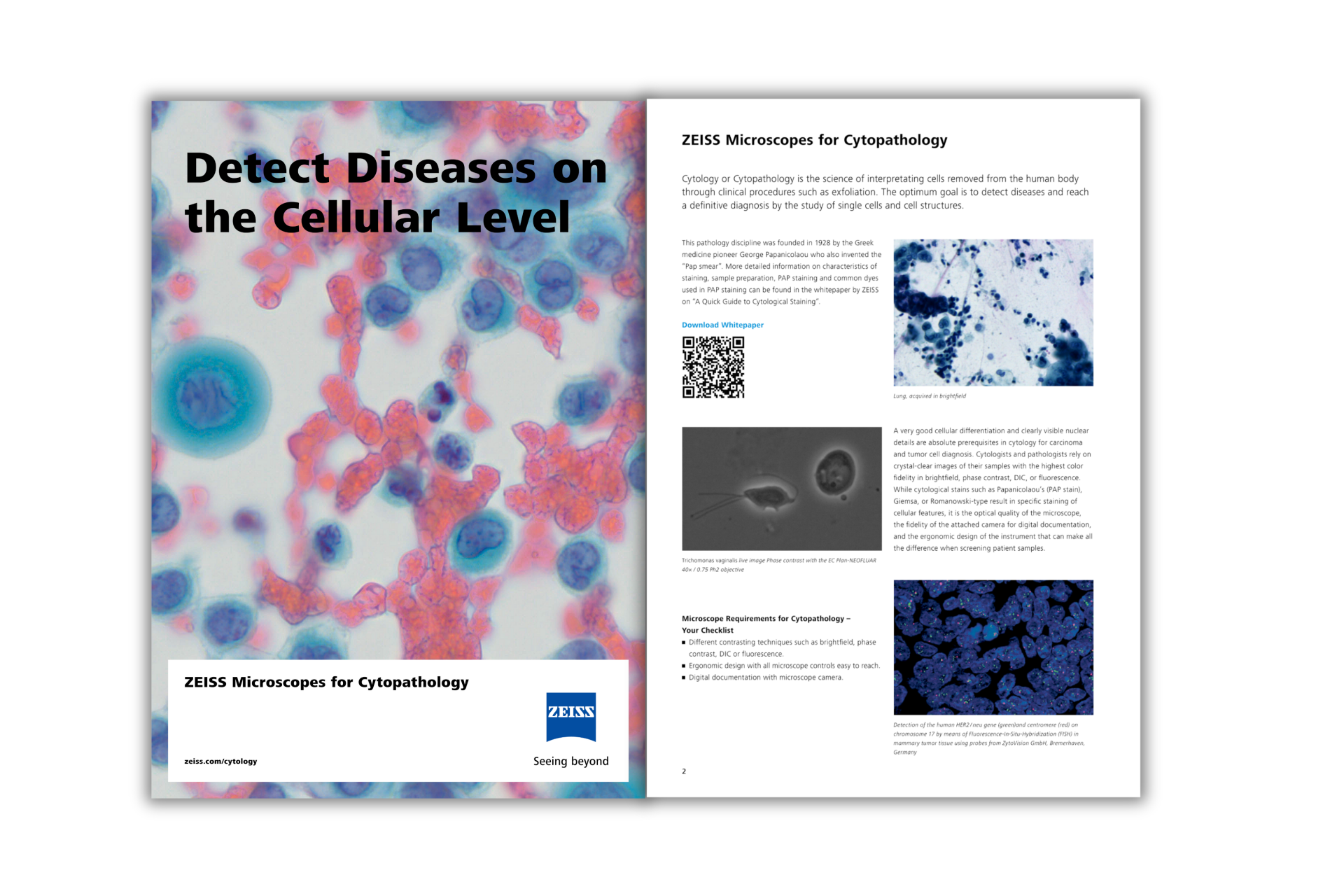
ZEISS Microscopy: Defining Excellence in Cytopathology
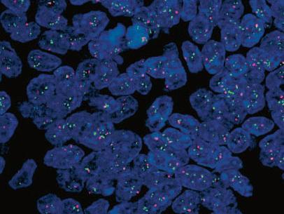
Cellular Diagnosis with Unmatched Details
As a cytopathologist, your work demands the highest level of cellular differentiation and accuracy. ZEISS understands these requirements. Our microscopes, equipped with cutting-edge technology, are designed to meet the intricacies of cytopathological analysis.
![]({"xsmall":"https://www.zeiss.com.sg/content/dam/rms/countries/singapore/apac/cyto-clear1.png/_jcr_content/renditions/original.image_file.100.75.0,0,807,605.file/cyto-clear1.png","small":"https://www.zeiss.com.sg/content/dam/rms/countries/singapore/apac/cyto-clear1.png/_jcr_content/renditions/original.image_file.360.270.0,0,807,605.file/cyto-clear1.png","medium":"https://www.zeiss.com.sg/content/dam/rms/countries/singapore/apac/cyto-clear1.png/_jcr_content/renditions/original.image_file.768.576.0,0,807,605.file/cyto-clear1.png","large":"https://www.zeiss.com.sg/content/dam/rms/countries/singapore/apac/cyto-clear1.png/_jcr_content/renditions/original.image_file.807.605.0,0,807,605.file/cyto-clear1.png","xlarge":"https://www.zeiss.com.sg/content/dam/rms/countries/singapore/apac/cyto-clear1.png/_jcr_content/renditions/original.image_file.807.605.0,0,807,605.file/cyto-clear1.png","xxlarge":"https://www.zeiss.com.sg/content/dam/rms/countries/singapore/apac/cyto-clear1.png/_jcr_content/renditions/original.image_file.807.605.0,0,807,605.file/cyto-clear1.png","max":"https://www.zeiss.com.sg/content/dam/rms/countries/singapore/apac/cyto-clear1.png/_jcr_content/renditions/original.image_file.807.605.0,0,807,605.file/cyto-clear1.png"})
![]({"xsmall":"https://www.zeiss.com.sg/content/dam/rms/countries/singapore/apac/cytofluorescence_her2-centromere_axiocam-mrm_2.jpg/_jcr_content/renditions/original.image_file.100.75.0,0,406,305.file/cytofluorescence_her2-centromere_axiocam-mrm_2.jpg","small":"https://www.zeiss.com.sg/content/dam/rms/countries/singapore/apac/cytofluorescence_her2-centromere_axiocam-mrm_2.jpg/_jcr_content/renditions/original.image_file.360.270.0,0,406,305.file/cytofluorescence_her2-centromere_axiocam-mrm_2.jpg","medium":"https://www.zeiss.com.sg/content/dam/rms/countries/singapore/apac/cytofluorescence_her2-centromere_axiocam-mrm_2.jpg/_jcr_content/renditions/original.image_file.406.305.0,0,406,305.file/cytofluorescence_her2-centromere_axiocam-mrm_2.jpg","large":"https://www.zeiss.com.sg/content/dam/rms/countries/singapore/apac/cytofluorescence_her2-centromere_axiocam-mrm_2.jpg/_jcr_content/renditions/original.image_file.406.305.0,0,406,305.file/cytofluorescence_her2-centromere_axiocam-mrm_2.jpg","xlarge":"https://www.zeiss.com.sg/content/dam/rms/countries/singapore/apac/cytofluorescence_her2-centromere_axiocam-mrm_2.jpg/_jcr_content/renditions/original.image_file.406.305.0,0,406,305.file/cytofluorescence_her2-centromere_axiocam-mrm_2.jpg","xxlarge":"https://www.zeiss.com.sg/content/dam/rms/countries/singapore/apac/cytofluorescence_her2-centromere_axiocam-mrm_2.jpg/_jcr_content/renditions/original.image_file.406.305.0,0,406,305.file/cytofluorescence_her2-centromere_axiocam-mrm_2.jpg","max":"https://www.zeiss.com.sg/content/dam/rms/countries/singapore/apac/cytofluorescence_her2-centromere_axiocam-mrm_2.jpg/_jcr_content/renditions/original.image_file.406.305.0,0,406,305.file/cytofluorescence_her2-centromere_axiocam-mrm_2.jpg"})
![]({"xsmall":"https://www.zeiss.com.sg/content/dam/rms/countries/singapore/apac/cytozeissaxiom-4.jpg/_jcr_content/renditions/original.image_file.100.75.0,1134,3023,3401.file/cytozeissaxiom-4.jpg","small":"https://www.zeiss.com.sg/content/dam/rms/countries/singapore/apac/cytozeissaxiom-4.jpg/_jcr_content/renditions/original.image_file.360.270.0,1134,3023,3401.file/cytozeissaxiom-4.jpg","medium":"https://www.zeiss.com.sg/content/dam/rms/countries/singapore/apac/cytozeissaxiom-4.jpg/_jcr_content/renditions/original.image_file.768.576.0,1134,3023,3401.file/cytozeissaxiom-4.jpg","large":"https://www.zeiss.com.sg/content/dam/rms/countries/singapore/apac/cytozeissaxiom-4.jpg/_jcr_content/renditions/original.image_file.1024.768.0,1134,3023,3401.file/cytozeissaxiom-4.jpg","xlarge":"https://www.zeiss.com.sg/content/dam/rms/countries/singapore/apac/cytozeissaxiom-4.jpg/_jcr_content/renditions/original.image_file.1280.960.0,1134,3023,3401.file/cytozeissaxiom-4.jpg","xxlarge":"https://www.zeiss.com.sg/content/dam/rms/countries/singapore/apac/cytozeissaxiom-4.jpg/_jcr_content/renditions/original.image_file.1440.1080.0,1134,3023,3401.file/cytozeissaxiom-4.jpg","max":"https://www.zeiss.com.sg/content/dam/rms/countries/singapore/apac/cytozeissaxiom-4.jpg/_jcr_content/renditions/original.image_file.1920.1440.0,1134,3023,3401.file/cytozeissaxiom-4.jpg"})
Do you face any of these issues at work?
- Are you encountering limitations in imaging quality during critical analyses?
- Do you require advanced staining solutions for nuanced cellular interpretation?
- Is ergonomic discomfort affecting your focus and productivity?
- Are you looking for more efficient digital integration in your workflow?
In this package, you will learn...
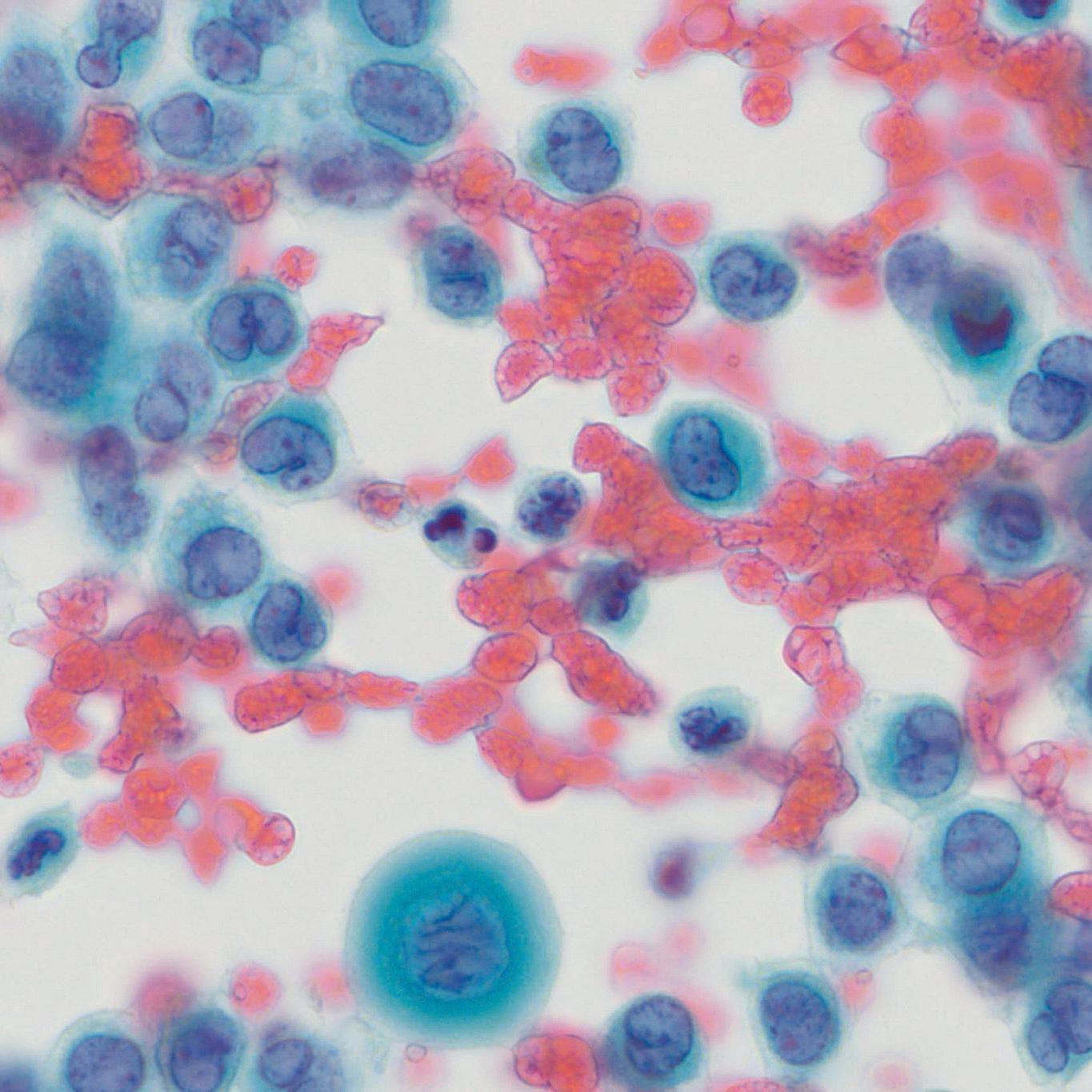
- Learn about the characteristics of staining including Papanicolaou (PAP) and Romanowsky stains , tailored for cytopathology.
- Learn how samples are prepared for cytological staining including the steps: Smear, Fixation, Permeabilization, Slide Mounting.
- Learn the principles of PAP staining including commonly used dyes, dye preparation and principles of staining
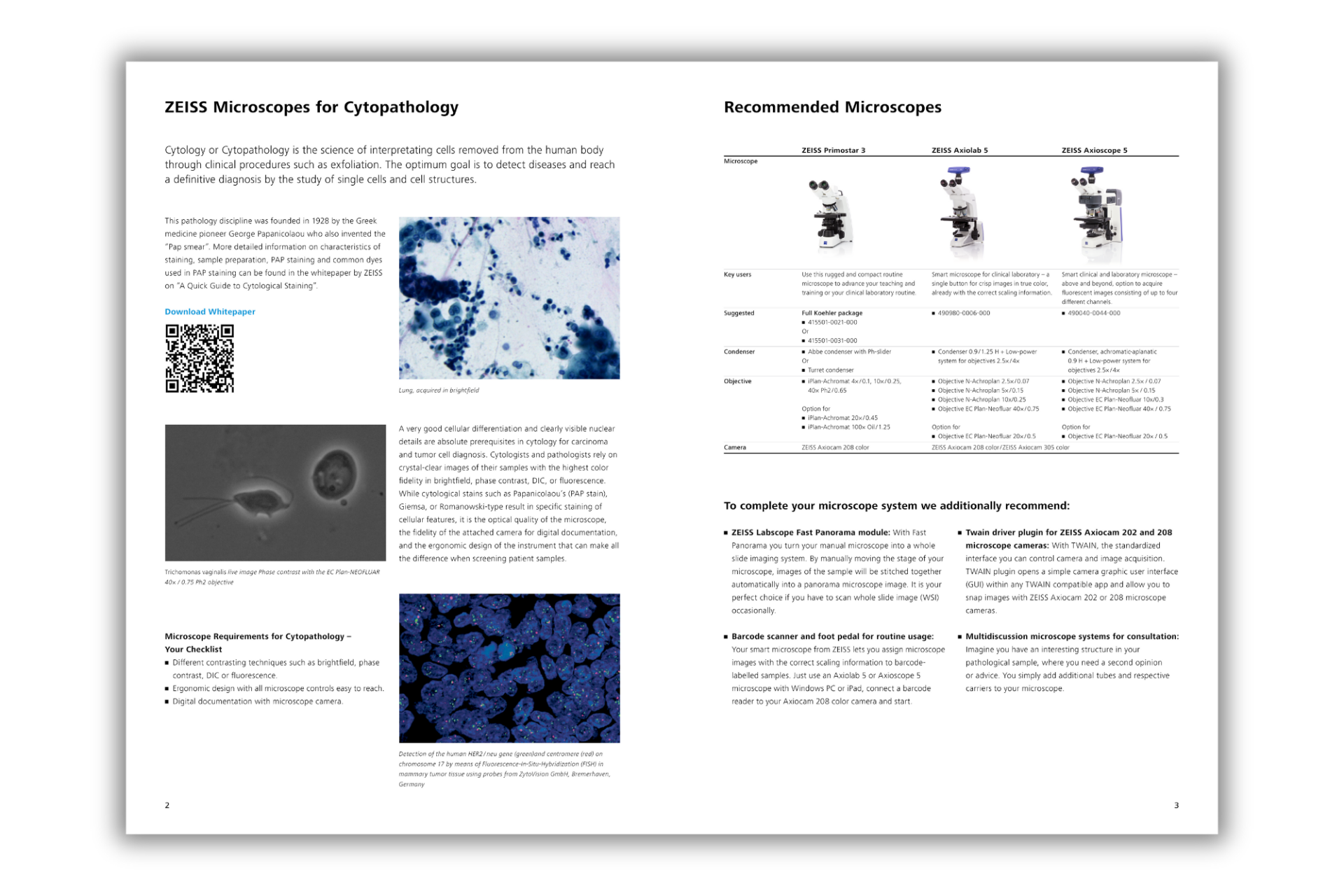
- Learn how the whole range of ZEISS microscopes cater to the complex needs of cytopathological analysis.
Here is what you will receive in the ZEISS Cytology Insights Package
- A Quick Guide to Cytological Staining (PDF)
- Quick Guide: ZEISS Microscopes for Cytopathology (PDF)